


Pros and Cons of Each and Why You Should Care
It’s no secret that adherent cells are more difficult to work with than suspension cells, but are they really just fussy?
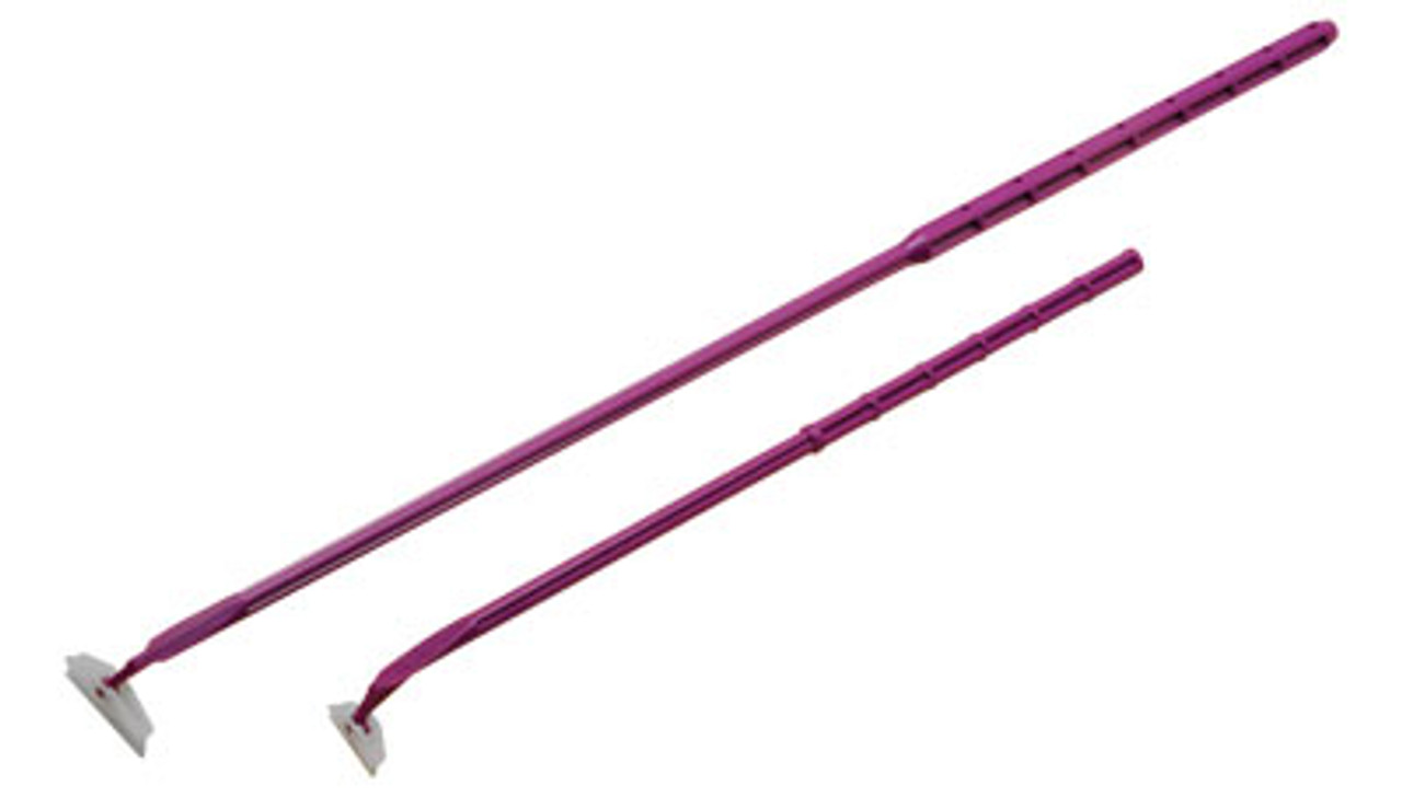
Pictured above; 25cm long cell scraper with rubber blade, sterile packed
This article discusses the two main approaches for replating adherent cultures. We’ll delve into what works best and how to troubleshoot.
Real science has no time for divas. Let’s get those cells in line!
Background on Trypsin
Trypsin is a pancreatic enzyme that is harvested for use in the enzymatic digestion of cell attachment proteins.
It is typically combined with the chelating agent EDTA to enhance its activity.
Pros and Cons
- Liquid, reaches corners easily
- Digests attachments over a wide range of concentrations
- Action is fast and uniform
- Somewhat gentler than mechanical removal
- Prone to contamination
- Expensive
- Changes cell protein composition and can release cells that don’t grow correctly
- Uses more materials
Spare the rod and spoil the culture, isn’t that what they say?
Background on Cell Scrapers
Cell scrapers are rubber-headed wands that are used to squeegee cells from their plates.
This method is actually as harsh as it seems! It does cause plasma membrane breakage and cell death.
Pros and Cons of Scraping
- Clean and simple
- Uses few materials
- Immediate results
- No troubleshooting hassles
- Harsh on weak and sickly cells
- Cells can break
- Caution to avoid contaminating or leaving cells behind
The major advantage is that some cells are better off dead than alive and not growing well on new plates.
Nature is tough sometimes, maybe that’s okay!
However, this IS why other methods are available for removing adherent cells from their containers.
Comparison Between Methods
Both are capable of killing cells, but there are benefits to each method that make them appealing.
Scrapers are cheap, easy to use, and disposable. There is virtually no preparation required before use.
The downside is that they are difficult to use on multiwell or smaller plates.
Trypsin is a powerful, uniform method for removing adherent cells. Because it is liquid, there is not as much risk of missing corners, and cells don’t stick or break as easily.
Regardless, it is costly and fully capable of destroying cells if misused.
Table Showing Relative Endpoint Success of Each Method
______________________________________________________________________
| Issue | Trypsin | Scraping |
| Cell breakage | Less than 10% for <1.5 min | 5-15% |
| Surface membrane protein changes | 10% | 36% |
_______________________________________________________________________________________
Studies have shown that both methods induce damage to the cell membrane.
This includes loss of membrane integrity and expression of proteins associated with cell death.
All-in-all, scraping is less notorious than enzymatic digestions are for changing protein expression in the long run.
It may be the more natural approach to replating cells, but not for endpoints if you don’t fix the cells first.
However, the takeaway here is that scraping can present fewer challenges for applications like FACS where surface antigens are viewed after, too.
Troubleshooting Common Issues
Scraping is less likely to intimidate new users than enzymatic digestion, but it’s an art. Knowing how to set up your workstation is key.
Some things to watch out for when you’re scraping cells:
- Low protein or nucleic acid yields
- Poor imaging with stains or dyes
- Cells not detaching easily
- Cells not growing on new plates
Things to try:
- Placing PBS and plates on ice while scraping
- Fixing cells in formalin before scraping
- Turning the plate more while scraping and performing an extra wash
- Scrape in warmed PBS or warmed media, check that everything is sterile
Trypsin can give the user an advantage because it works well at a fairly wide range of concentrations, but 0.25% is the established working one.
Some things to watch out for:
- Low protein yield or nucleic acid yield
- Poor imaging results
- Cells not detaching quickly enough
- Contamination
- Poor regrowth
Try doing this:
- Optimize concentrations so you use the lowest one with the fastest results
- Try raising the concentration or extending the time if cells are not detaching
- Incubate the cells for up to five minutes at the most
- Make sure everything is filtered for use in cell culture beforehand
- Deactivate trypsin with FBS before removing it from the cells
Stellar Bargains on Cell Culture Consumables
Stellar Scientific carries a selection of scrapers for cell culture. Whether you need a pivoting head, a traditional one, or an extra long handle, we’ve got it covered.
Browse our catalog and choose from all of the finest sterile packed, RNASE and DNASE free lab consumables at great prices.
If you aren’t finding what you need, don’t hesitate to reach out to us through our contact us page!
Footnotes:
__________________
- Huang, H. L., Hsing, H. W., Lai, T. C., Chen, Y. W., Lee, T. R., Chan, H. T., Lyu, P. C., Wu, C. L., Lu, Y. C., Lin, S. T., Lin, C. W., Lai, C. H., Chang, H. T., Chou, H. C., & Chan, H. L. (2010). Trypsin-induced proteome alteration during cell subculture in mammalian cells. Journal of biomedical science, 17(1), 36. https://doi.org/10.1186/1423-0127-17-36
- Lordon, Blandine, et al. “Impact of trypsin on cell cytoplasm during detachment of cells studied by terahertz sensing.” Biophysical Journal, June 2024, https://doi.org/10.1186/1423-0127-17-36
- Nowak-Terpiłowska, A., Śledziński, P., & Zeyland, J. (2021). Impact of cell harvesting methods on detection of cell surface proteins and apoptotic markers. Brazilian journal of medical and biological research = Revista brasileira de pesquisas medicas e biologicas, 54(2), e10197. https://doi.org/10.1186/1423-0127-17-36
- YAN, GE, and THOMAS EFFERTH. “Cell harvesting methods affect cellular integrity of adherent cells during apoptosis detection.” Anticancer Research, vol. 38, no. 12, 30 Nov. 2018, pp. 6669–6672, https://doi.org/10.1186/1423-0127-17-36