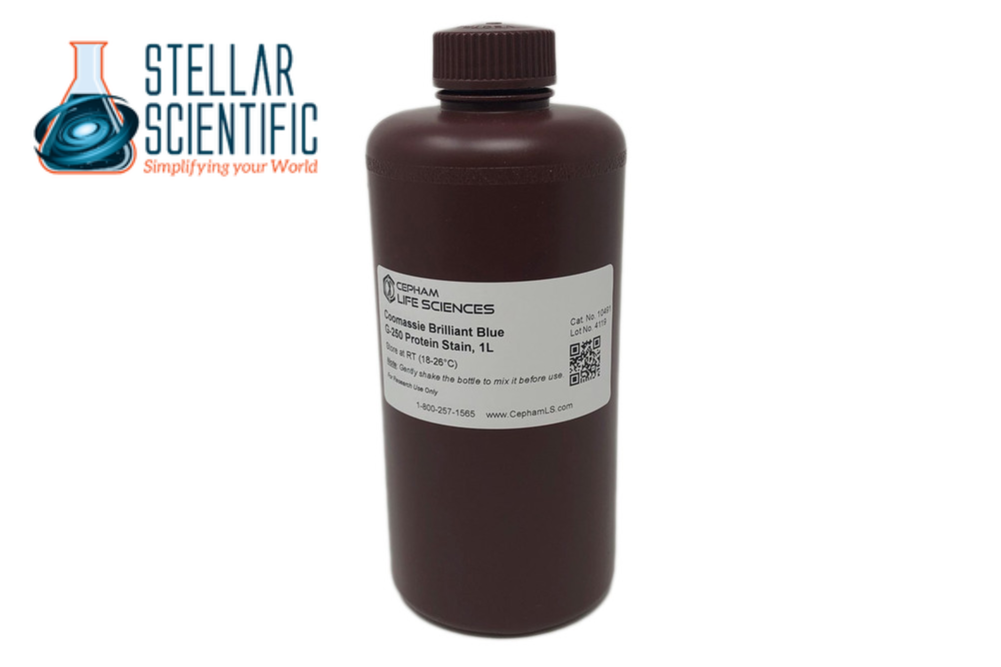
In the world of scientific research, particularly in molecular biology and biochemistry, detecting and quantifying proteins is crucial. Proteins are the building blocks of life, involved in countless biological processes, and understanding their behavior is key to advancing medical, environmental, and pharmaceutical research. One of the most widely used methods for detecting proteins in the laboratory is Coomassie Blue staining. This technique has proven to be a reliable and effective way to visualize and quantify proteins in complex biological samples.
In this blog post, we’ll explore how Coomassie Blue works, its applications in protein detection, and why it remains one of the most popular techniques in laboratories worldwide.

What is Coomassie Blue?
Coomassie Blue is a dye that binds to proteins, turning them blue and allowing them to be easily visualized under specific light conditions. The dye works by interacting with the amino acid side chains, especially those of arginine, histidine, and phenylalanine, which are abundant in proteins. When the dye binds to proteins, it forms a stable complex that shifts in color, typically from brown to blue, which can then be detected visually or quantified.
There are two main forms of Coomassie Blue dye: Coomassie Brilliant Blue G-250 and Coomassie Brilliant Blue R-250. The G-250 form is typically used for protein quantification, while R-250 is often used for gel staining, such as in SDS-PAGE (sodium dodecyl sulfate-polyacrylamide gel electrophoresis).
How Coomassie Blue Works
The principle behind Coomassie Blue protein detection is relatively simple. Here’s how the process works:
- Preparation of the Sample: Proteins are typically separated using electrophoresis (such as SDS-PAGE) or prepared as a solution. The sample can be loaded into a gel or placed in a well for direct analysis.
- Staining: After the proteins are separated or in solution, Coomassie Blue dye is added to the sample. The dye binds to the proteins in the sample, causing a color change to blue. The degree of color intensity corresponds to the amount of protein present.
- Visualization: The stained proteins can be visualized directly on a gel or membrane using simple techniques like light absorption or photography. In some cases, the sample may undergo destaining, where excess dye is removed, and only the protein-bound dye remains.
- Quantification: For quantification purposes, spectrophotometry can be used to measure the absorbance of the stained proteins at specific wavelengths (usually 595 nm for Coomassie Brilliant Blue). The intensity of the color correlates with the concentration of protein in the sample.
Applications of Coomassie Blue in Protein Detection
Coomassie Blue stain has widespread applications in both protein quantification and protein visualization, making it an essential tool in many areas of scientific research.
1. Protein Quantification
One of the primary uses of Coomassie Blue is in the quantification of proteins. Researchers often need to know the concentration of a protein in a sample for further analysis, and Coomassie Blue provides an easy and reliable way to do this. The amount of protein can be quantified by comparing the absorbance of the stained sample to a standard curve generated from known concentrations of protein.
This is particularly useful in cases where other methods like Bradford assays or BCA assays may be too complex or time-consuming. Coomassie Blue provides a straightforward, cost-effective method for determining protein concentrations in a variety of sample types.
2. SDS-PAGE Gel Staining
In SDS-PAGE, proteins are separated based on their size by applying an electric field to a gel. After electrophoresis, the gel is often stained with Coomassie Blue to visualize the separated protein bands. This allows researchers to analyze the size and abundance of proteins present in the sample.
Coomassie Blue is one of the most common stains used for SDS-PAGE gels because it is highly sensitive and provides clear, sharp bands for easy identification. It is also relatively quick and simple, making it an ideal choice for labs performing routine protein analysis.
3. Western Blotting
After proteins are separated via SDS-PAGE, they are often transferred to a membrane for Western blotting, where specific proteins are detected using antibodies. While Coomassie Blue is not used directly for antibody-based detection, it can be used as a reference for the total protein content in the sample. By staining the membrane with Coomassie Blue before performing a Western blot, researchers can ensure that the amount of protein loaded into each lane is consistent.
4. Protein Purification
Coomassie Blue is also used during protein purification protocols to assess the purity of a protein sample. When proteins are purified, researchers often need to determine how much protein is present and whether it is the correct protein of interest. Coomassie Blue staining can help visualize the purity of the sample by showing the presence of contaminants or confirming the successful isolation of a single protein band.
5. Cellular Protein Analysis
In cell biology, Coomassie Blue is used to assess the protein content of cell lysates or tissue extracts. By analyzing the protein profile of cells or tissues, researchers can gain insights into the biological processes at play. Coomassie Blue staining can help identify changes in protein expression levels due to experimental treatments or cellular stress.

Advantages of Coomassie Blue
1. Simplicity and Cost-Effectiveness
One of the biggest advantages of using Coomassie Blue is its simplicity. The staining process is straightforward and does not require specialized equipment or reagents. Additionally, it is an affordable option compared to other more complex and expensive protein detection methods like mass spectrometry or radioactive labeling.
2. Sensitivity
Coomassie Blue is highly sensitive, allowing researchers to detect even low amounts of protein in a sample. This sensitivity makes it suitable for a wide range of protein concentrations and applications, from small-scale experiments to large-scale protein assays.
3. Rapid Results
Coomassie Blue staining typically provides fast results, which is important for many researchers working under time constraints. Staining and visualization can often be completed in less than an hour, which is particularly useful in high-throughput settings or when working with large numbers of samples.
4. Broad Applicability
Coomassie Blue is versatile and can be used in a variety of formats, from gels and blots to liquid samples. Its broad applicability makes it an ideal choice for many types of protein analysis.
Limitations of Coomassie Blue
While Coomassie Blue is widely used, it does have some limitations. For example, it may not be as sensitive as other protein assays, such as fluorescent-based methods or radioactive assays, especially at very low protein concentrations. Additionally, it can sometimes produce background staining or interfere with downstream analyses if not properly washed or destained.
About Stellar Scientific
At Stellar Scientific, we are committed to providing high-quality laboratory equipment and reagents for researchers in various fields, including protein analysis and biochemical research. Our products, including Coomassie Blue dye, are designed to support your research needs and help you achieve accurate, reliable results. We offer a range of laboratory tools and equipment, from microplate readers to orbital shakers, to ensure that your experiments are efficient and effective.


