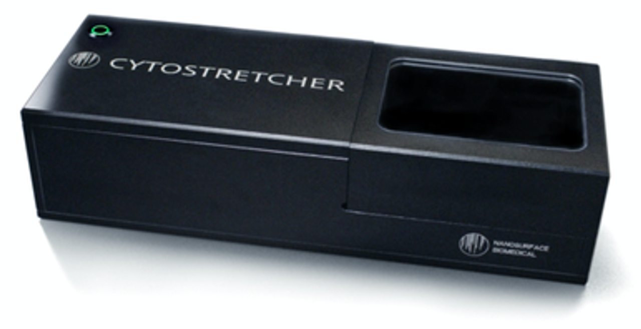
Stellar Scientific is honored to have supplier partners like NanoSurface Biomedical that make time to share their knowledge with us and our customers.
What is a cytostretcher, or cell stretcher, and how is it useful for laboratories that grow cells?
The article below explains the impact mechanical stress plays on cellular growth and how a cytostretcher adds complexity to cell growth.
Every day we learn more about the importance of the relationship between the cell and its environment.
Though most cell biology is performed on glass or plastic petri dishes and culture flasks, it turns out that many cells need specific cues from their environment to grow, develop, and function properly.
These cues come from the extracellular matrix, or ECM.
First, specific shape of the ECM plays an important role in determining cell and tissue shape.
For example, skeletal 1,2 and heart3 muscle cells grow in a linear mesh of collagen and fibronectin which guides them into a laminar orientation that synchronizes the direction of their contraction, making coordinated movements possible.
Second, the molecules that make up the ECM that surrounds cells are important too 4,5. While proteins like collagen, fibronectin, and laminin all play a role in modulating cell processes, these different proteins have unique roles in regulating distinct pathways 6.
Last, it turns out that mechanical stretch plays a role in the proper development and regulation of cells and tissues.
Cells can feel mechanical stress and use that to change gene transcription 7 and protein expression8,9 pathways. Mechanical stress can even activate ion channels to produce immediate responses 10,11.
Understanding how externally-applied mechanical stress is important to researchers working across all the organs of the body. From the brain 12, bone13,14, skin15, eyes16, heart3,17,18, blood vessels19,20, and liver21, mechanical stress is ubiquitous across the body.
For example, a healthy human heart stretches about 5-10% per beat 22–24.
Applying this kind of stretch to cells in culture can re-organize their structure and can stimulate the expression of normal, healthy genes.
Indeed, healthy mechanical stretch directs the organization of the cytoskeleton and of healthy intracellular gap junctions.
Diseases like high blood pressure can increase stretch above this threshold, resulting in the remodeling of the muscle tissue and expression of several undesirable genes 25,26.
In addition to cyclical stretch, some organs require variable stretch to maintain healthy phenotypes.
For example, a healthy human lung has a variable rate of stretch throughout the day.
It has been shown that lung cells that are given a variable stretch routine show highly reduced markers of inflammation compared to those that just get a constant, cyclical stretch 27.
NanoSurface Biomedical’s Cytostretcher was designed to integrate all of these extracellular cues in an instrument that gives you unparalleled flexibility to apply different stretch routines.
The Cytostretcher chambers use a proprietary patterned chamber that simulates the shape of the cell’s ECM and can be functionalized with a variety of ECM proteins.
Stretch routines are designed with the Cytostretcher’s easy to use software, which enables you to make your own custom stretch routines with no programming skills required.
Mechanical stress can be applied to cells in the culture incubator with the NanoSurface Cytostretcher or on the optical microscope stage for simultaneous observation using the Cytostretcher-LV.
A variety of examples are shown below.
| Tissue | Magnitude | Rate |
| Lung; healthy 27 | Vary randomly between 1-15% | 0.5Hz |
| Lung; inflammatory response 27 | 7.5% | 0.5Hz |
| Healthy Heart 22–24 | 5-10% | 0.5-2Hz |
| Unhealthy Heart 28,29 | >12% | >2Hz |
| Blood Vessel: healthy vascular Endothelial 30,31 | 8-10% | 0.1-1Hz, cyclical |
| Skeletal Muscle: increase glucose metabolism 32,33 | 20% | 0.25Hz for 60s, 30m rest, repeat. |
| Eye: Age-related macular degeneration 34 | 25% | 0.125Hz for 20-24h |
| Neural Stem cell: traumatic injury 35 | >38% | Single Ramp |
In addition to the Cytostretcher, Nanosurface Bio has developed a line of nano-patterned cell culture ware which are now available in single 35mm dish, 24 and 96 well plate formats.
Unlike flat polystyrene cell culture dishes, flasks and plates, the nano-patterned cell culture surface provides tiny grooves that let cells pile up and align more closely to how they operate in-vivo.
Whether you need routine plastic lab consumables for your cell culture or more specialized cell culture plastics, Stellar Scientific offers a robust variety of products for most every lab.
Footnotes:
1. Bettadapur, A. et al. Prolonged Culture of Aligned Skeletal Myotubes on Micromolded Gelatin Hydrogels. Sci. Rep. 6, (2016).
2. Provenzano, P. P. & Vanderby, R. Collagen fibril morphology and organization: Implications for force transmission in ligament and tendon. Matrix Biol. 25, 71–84 (2006).
3. Kim, D.-H. et al. Nanoscale cues regulate the structure and function of macroscopic cardiac tissue constructs. Proc. Natl. Acad. Sci. 107, 565–570 (2010).
4. Järveläinen, H. Extracellular matrix molecules: potential targets in pharmacotherapy. Pharmacol Rev. 61, 198–223 (2009).
5. Bonnans, C., Chou, J. & Werb, Z. Remodelling the extracellular matrix in development and disease. Nat. Rev. Mol. Cell Biol. 15, 786–801 (2014).
6. Gattazzo, F., Urciuolo, A. & Bonaldo, P. Extracellular matrix: A dynamic microenvironment for stem cell niche. Biochim. Biophys. Acta - Gen. Subj. (2014).
7. Tajik, A. et al. Transcription upregulation via force-induced direct stretching of chromatin. Nat. Mater. 15, 1287–1296 (2016).
8. Wei, S. C. et al. Matrix stiffness drives epithelial-mesenchymal transition and tumour metastasis through a TWIST1-G3BP2 mechanotransduction pathway. Nat. Cell Biol. 17, 678–688 (2015).
9. Hu, B. et al. Mechanical stretch suppresses microRNA-145 expression by activating extracellular signal-regulated kinase 1/2 and upregulating angiotensin- converting enzyme to alter vascular smooth muscle cell phenotype. PLoS One 9, 1–10 (2014).
10. Wu, J., Lewis, A. H. & Grandl, J. Touch, Tension, and Transduction – The Function and Regulation of Piezo Ion Channels. Trends Biochem. Sci. 42, 57–71 (2017).
11. Ranade, S. S., Syeda, R. & Patapoutian, A. Mechanically Activated Ion Channels. Neuron 87, 1162–1179 (2015).
12. Christopherson, G. T., Song, H. & Mao, H. Q. The influence of fiber diameter of electrospun substrates on neural stem cell differentiation and proliferation. Biomaterials 30, 556–564 (2009).
13. Engler, A. J., Sen, S., Sweeney, H. L. & Discher, D. E. Matrix Elasticity Directs Stem Cell Lineage Specification. Cell 126, 677–689 (2006).
14. Rosa, A. L. et al. Nanotopography drives stem cell fate toward osteoblast differentiation through ??1??1 integrin signaling pathway. J. Cell. Biochem. 115, 540–548 (2014).
15. Wang, J. et al. An updated review of mechanotransduction in skin disorders: Transcriptional regulators, ion channels, and microRNAs. Cell. Mol. Life Sci. 72, 2091–2106 (2015).
16. Raghunathan, V. K. et al. Role of substratum stiffness in modulating genes associated with extracellular matrix and mechanotransducers YAP and TAZ. Investig. Ophthalmol. Vis. Sci. 54, 378–386 (2013).
17. Ruan, J. L. et al. Mechanical Stress Conditioning and Electrical Stimulation Promote Contractility and Force Maturation of Induced Pluripotent Stem Cell-Derived Human Cardiac Tissue. Circulation 134, 1557–1567 (2016).
18. Qiu, Y. et al. A role for matrix stiffness in the regulation of cardiac side population cell function. Am. J. Physiol. Circ. Physiol. 308, H990–H997 (2015).
19. Hahn, C. & Schwartz, M. A. Mechanotransduction in vascular physiology and atherogenesis. Nat. Rev. Mol. Cell Biol. 10, 53–62 (2009).
20. Lehoux, S. & Tedgui, A. Cellular mechanics and gene expression in blood vessels. J. Biomech. 36, 631–643 (2003).
21. Saneyasu, T., Akhtar, R. & Sakai, T. Molecular Cues Guiding Matrix Stiffness in Liver Fibrosis. Biomed Res. Int. 2016, (2016).
22. Mihic, A. et al. The effect of cyclic stretch on maturation and 3D tissue formation of human embryonic stem cell-derived cardiomyocytes. Biomaterials 35, 2798–2808 (2014).
23. Zimmermann, W. H. et al. Tissue engineering of a differentiated cardiac muscle construct. Circ Res 90, 223–230 (2002).
24. MacQueen, L. A. et al. A tissue-engineered scale model of the heart ventricle. Nat. Biomed. Eng. (2018). doi:10.1038/s41551-018-0271-5
25. McCain, M. L., Sheehy, S. P., Grosberg, A., Goss, J. A. & Parker, K. K. Recapitulating maladaptive, multiscale remodeling of failing myocardium on a chip. Proc. Natl. Acad. Sci. (2013). doi:10.1073/pnas.1304913110
26. Onodera, T., Tamura, T., Said, S., McCune, S. A. & Gerdes, A. M. Maladaptive remodeling of cardiac myocyte shape begins long before failure in hypertension. Hypertension 32, 753–757 (1998).
27. Santos, L. et al. Variable stretch reduces the pro- inflammatory response of alveolar epithelial cells. 1–16 (2017). doi:10.1371/journal.pone.0182369
28. Saffitz, J. E. & Kléber, A. G. Effects of mechanical forces and mediators of hypertrophy on remodeling of gap junctions in the heart. Circ Res 94, 585–591 (2004).
29. Gopalan, S. M. et al. Anisotropic stretch-induced hypertrophy in neonatal ventricular myocytes micropatterned on deformable elastomers. Biotechnol Bioeng 81, 578–587 (2003).
30. Birukov, K. G. Cyclic Stretch, Reactive Oxygen Species, and Vascular Remodeling. Antioxid. Redox Signal. 11, 1651–1667 (2009).
31. Greiner, A. M., Biela, S. A., Chen, H., Spatz, J. P. & Kemkemer, R. Featured Article: Temporal responses of human endothelial and smooth muscle cells exposed to uniaxial cyclic tensile strain. Exp. Biol. Med. 240, 1298–1309 (2015).
32. Passey, S., Martin, N., Player, D. & Lewis, M. P. Stretching skeletal muscle in vitro: Does it replicate in vivo physiology? Biotechnol. Lett. 33, 1513–1521 (2011).
33. Hatfaludy, S., Shansky, J. & Vandenburgh, H. H. Metabolic alterations induced in cultured skeletal muscle by stretch-relaxation activity. Am. J. Physiol. 256, C175-81 (1989).
34. Wu, S. et al. Cyclic stretch induced-retinal pigment epithelial cell apoptosis and cytokine changes. BMC Ophthalmol. 17, 1–9 (2017).
35. Sherman, S. A. et al. Stretch Injury of Human Induced Pluripotent Stem Cell Derived Neurons in a 96 Well Format. Sci. Rep. 6, 1–12 (2016).


