The wizardry of flow cytometry is breaking new ground in molecular biology, protein engineering, pathology, immunology and other research disciplines.
Using laser light, reagents and fluorophores, researchers can analyze tens of thousands of cells quickly and develop a clear picture of the different components and attributes of the cells they are trying to better understand.
As its name implies, the flow cytometer differs from standard microscopy in that cellular analysis takes place while a stream of cells flows through the device and not in a contained flask, plate of dish.
To achieve a smooth flow of cells, it is important to properly prepare the sample. The type of cells to be examined and other potential impediments that can disrupt the flow need to be considered.
For example: Most flow cytometers come with different nozzles to channel the flow of cells based on size and type. Lymphocytes and other small cells use the smaller 70 micron tip while adherent cell lines require the larger 100 micron tip.
The cell sample should not contain too high concentration of dead cells which can release soluble DNA that will coat the remaining cells and cause clumping.
Similarly, adherent cells, because of their natural clinging tendency, can join together and clog the flow cytometer nozzle.
Proper preparation often involves running the sample first through a cell-strainer; basically a piece of mesh with a specific porosite size to create a single-cell suspension.
The most common mesh sizes for cell strainers are 40, 70 and 100 microns.
For nearly a generation, the design and function of the cell strainer has remained essentially the same.
A polypropylene basket with mesh panels on the bottom and surrounding sides is inserted into a 50mL conical tube and the cell suspension is poured or pipetted through.
To help preserve sterility the cell strainer has a thumb-tab that allows for manipulation without coming in contact with the strainer itself.

While effective, the classic version of the cell strainer presents several challenges:
1. The mesh on the sides of the strainer press up against the walls of the conical tube and serve no actual function.
2. The basket design allows for air pressure inside the conical tube to create a "blow-back" effect that blocks the liquid from flowing through.
3. Classic cell strainers are uniformly designed to fit 50mL tubes. When preparing a suspension of 14mL or less, a lot of plastic is wasted unnecessarily.
4. In situations where serial straining is required (say, first a 100um followed by a 70um), two 50mL conical tubes must be used.
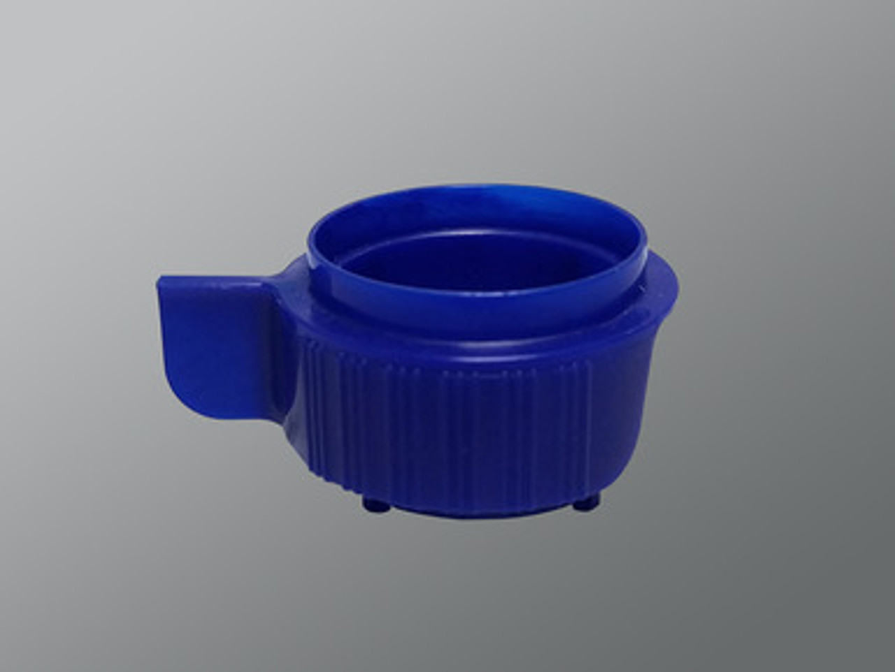
Stellar Scientific introduces the SWiSH™ brand of cell strainers with unique features that address these concerns.

Available color-coded in 40, 70 and 100um size, SWiSH™cell strainers have done away with side mesh and offer a body redesign that results in a sloped and shapely form.
Together, this produces a more rapid and even flow as is evidenced by this short video presentation:
SWiSH™cell strainers are stackable for serial straining
SWiSH™cell strainers can be fitted with the optional reducing adapter that is compatible with 15mL, 5mL and FACS tubes, reducing laboratory waste and facilitating faster prep time.

If you would like to blow through your flow cytometry sample prep, ask us for a free sample of the SWiSH™cell strainer and reducing adapter.
We think they will "flow your mind!"


