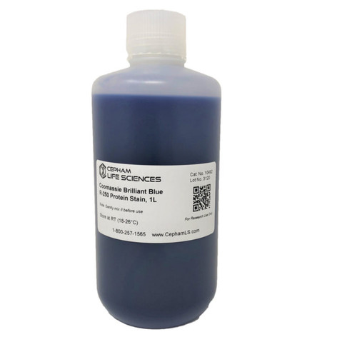
The sensitivity of our Coomassie Brilliant Blue G‐250 Protein Stain is up to 8 ng protein/ band, as observed in 4‐20% SDS-PAGE gel loaded with BSA.
Coomassie Brilliant Blue G‐250 Protein Stain binds to proteins through ionic interactions between the sulfonic acid groups of the dye and positive protein amine groups and through Van der Waals attractions.
Coomassie Brilliant Blue G‐250 Protein Stain can be used with SDS-PAGE, Native PAGE, Tricine gels with fixation step, 2-D Electrophoresis, etc
The capability of G-250 to create a rapid and convenient staining procedure is due to its particular properties and manifests a leuco form below pH 2.
Solutions of the dye, dark blue black at pH 7, turn a clear tan upon acidification. The leuco form recovers its blue color upon binding to protein, apparently due to the more neutral pH of the environment around the protein molecule.
Under ideal staining conditions, a gel placed in Coomassie Brilliant Blue G-250 Stain will manifest blue protein bands on a light amber background. The protein bands develop rapidly and there is no need to de-stain, as the background color is not dark.
Coomassie Brilliant Blue G‐250 stains the proteins with high band visibility and the protein bands up to 0.1 µg protein can be observed within 5 minutes, while staining the gel.
Features:
- Ready to use solution
- Fast protein staining – in about 60-90 minutes
- Detects up to 8 ng protein per band
- Protein bands appear and visible while the gel in stain
- Compatible with fixing by alcohol
- Mass Spec compatible


